1
Upload medical images
More than 10 services for different modalities
Mammograms, radiographs, fluorograms, CT, MRI, CBCT, skin images, and even OCT
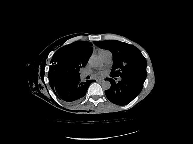
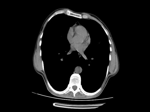
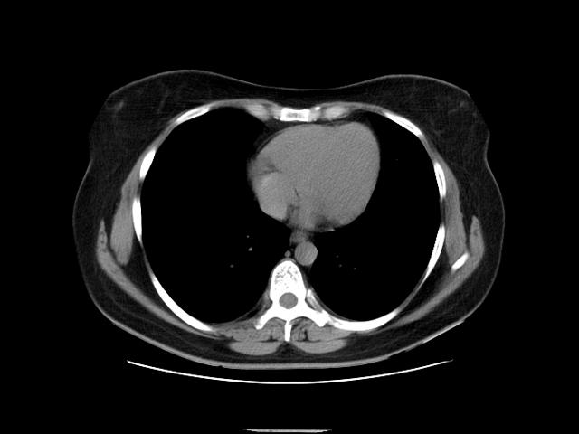
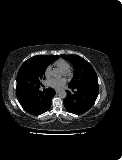
AI service for the detection and quantitative assessment of pathologies of the lungs, mediastinum, and bony structures of the spine
Upload DICOM files and view them immediately in your browser
More than 10 services for different modalities
Mammograms, radiographs, fluorograms, CT, MRI, CBCT, skin images, and even OCT
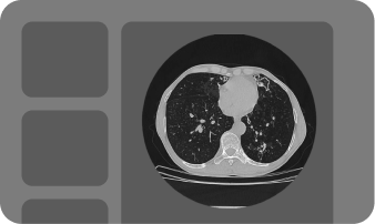
Upload studies in DICOM format; AI removes personal data
The service automatically measures standard parameters, while you work with the images in a familiar viewer, as you prefer
AI will describe the study — all you have to do is double-check it
Processed studies and the report can be downloaded
Identifies and quantitatively digitizes predictors of cardiovascular disease: coronary artery calcification, pulmonary trunk enlargement, thoracic aortic dilatation, paracardiac fat, pericardial effusion, and hydrothorax.
The physician receives a separate cardiac series for rapid patient screening.
Automatically performs routine measurements and calculates key quantitative parameters.
In the completed examination report, the physician receives the sizes and volumes of pulmonary lesions, the diameters of the thoracic aorta and pulmonary trunk, the volume of pleural and pericardial effusion, the volume of paracardiac fat, the pneumothorax volume, the percentage of emphysema, the Agatston score, the CAC-DRS category, and separate calcium scores for each artery.
Visualizes the results of AI analysis in lung and soft-tissue windows, helping the physician assess changes in the lungs, pleura, heart, and major vessels without the need to switch between separate results.
Automatically generates a curved planar reformation of the spine, allowing the physician to assess the vertebrae in a single image and identify compression deformities more quickly.
An intelligent assistant that can answer questions using an extensive medical library.