1
Upload medical images
More than 10 services for different modalities
Mammograms, radiographs, fluorograms, CT, MRI, CBCT, skin images, and even OCT
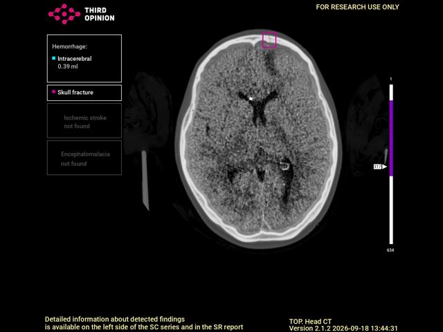
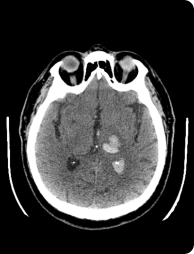
AI service for diagnosing ischemic stroke, intracranial hemorrhages, and skull fractures, as well as for automating morphometry
Upload DICOM files and view them immediately in your browser
More than 10 services for different modalities
Mammograms, radiographs, fluorograms, CT, MRI, CBCT, skin images, and even OCT
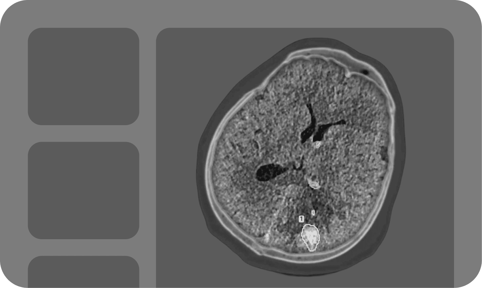
Upload studies in DICOM format; AI removes personal data
The service automatically measures standard parameters, while you work with the images in a familiar viewer as you prefer
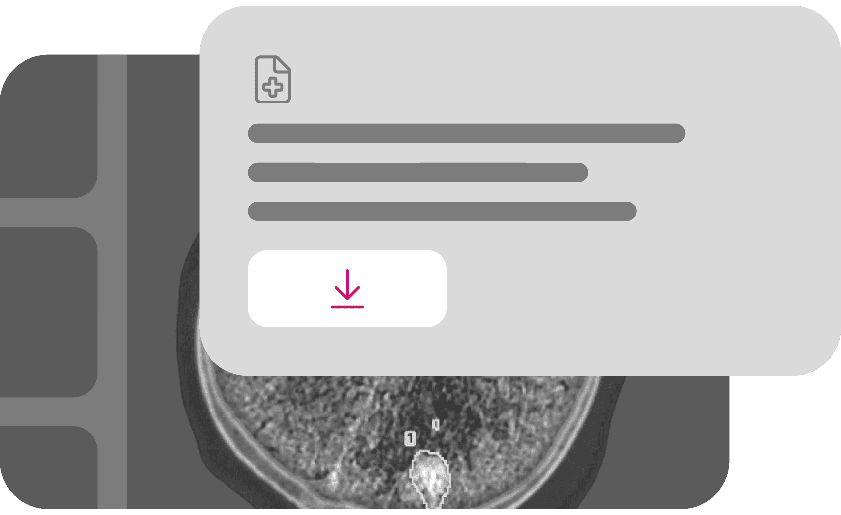
AI will describe the study—you will only need to verify it
Processed studies and the report can be downloaded
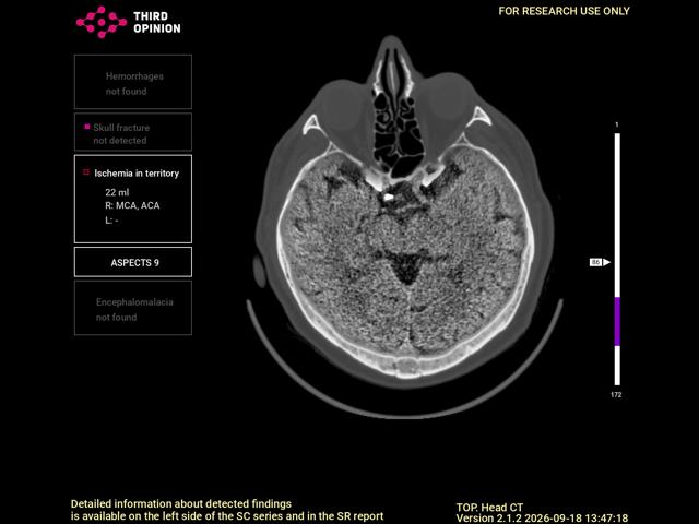
Segments the ischemic area, determines its volume and the affected vascular territory
Automatically calculates ASPECTS with differentiation of chronic changes, reducing the amount of manual measurements.
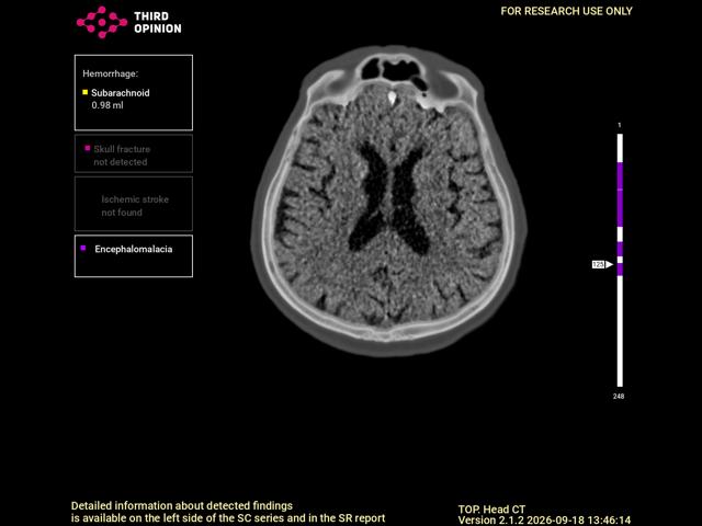
Identifies the main types of intracranial hemorrhage and calculates the volume of each lesion.
Quantitative data help the physician assess the patient's condition and choose further management.
Detects fractures of the cranial vault, skull base, and facial bones.
Visualization of the detected changes helps the physician evaluate studies of patients with traumatic brain injury.
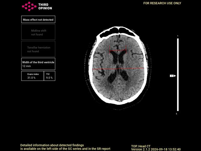
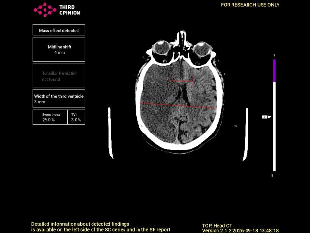
Automatically measures midline shift, the degree of cerebellar tonsillar herniation, the width of the third ventricle, and ventriculocranial indices.
Quantitative measures make it possible to monitor changes without manual measurements.
An intelligent assistant that can answer questions using an extensive medical library.